
BLOG
Besos Meaning: A Journey into the Heart of Latin American Culture

Besos is derived from the Spanish language, where it translates to “kisses.” However, the meaning of besos goes beyond a simple translation. It’s a term that encompasses a range of emotions, from affection and love to passion and intimacy.
The Cultural Significance of Besos
In many Latin American cultures, besos are an integral part of daily life. They’re a way to show affection, greet one another, and express love. The cultural significance of besos is deeply rooted in the values of warmth, hospitality, and closeness.

The Different Types of Besos
While the term “besos” is often associated with romantic love, it’s not the only context in which it’s used. Besos can be exchanged between family members, friends, and even as a greeting or farewell.
- A beso on the cheek is a common greeting in many Latin American countries.
- A beso on the lips is often reserved for romantic partners or loved ones.
- A beso on the forehead or hand can be a sign of respect, affection, or blessing.
“I remember my abuela giving me besos on the forehead every night before bed. It was a special moment that made me feel loved and safe.”
The Emotional Significance of Besos
Besos are more than just a physical gesture; they’re a way to convey emotions and connect with others. The act of giving or receiving a beso can evoke feelings of comfort, security, and love.
FAQs
Q: What is the meaning of besos in Spanish?
A: Besos is the Spanish word for “kisses.” It’s a term used to describe a range of affectionate gestures, from romantic kisses to friendly pecks on the cheek.
Q: How do you use besos in a sentence?
A: You can use besos in a sentence to express affection or love, such as “Dale besos a tu familia de mi parte” (Give your family a kiss from me).
Q: What is the cultural significance of besos in Latin America?
A: Besos play a significant role in Latin American culture, where they’re used to show affection, greet one another, and express love.
Q: Can besos be used in non-romantic contexts?
A: Yes, besos can be used in non-romantic contexts, such as between family members or friends. It’s a way to show affection and closeness.
Conclusion
Besos, we discover that it’s more than just a word – it’s a way to connect with others and express our emotions. Whether you’re looking to deepen your understanding of Latin American culture or simply want to show affection to those around you, besos is a term that’s worth exploring further.

On June 28, 2025, Air France flight AF136 operated by an Airbus A350-900 departed Paris-Charles de Gaulle (CDG) bound for Chicago O’Hare International Airport (ORD). What should have been a routine transatlantic crossing turned into an extraordinary nine-hour ordeal when the aircraft was denied landing authorization at its destination, forcing it to turn back to Paris mid-Atlantic.
This article provides a comprehensive breakdown of the incident: a detailed timeline, the potential reasons behind the denied clearance, what happened to the affected passengers, and what rights they are entitled to under EU law. It also examines Air France’s use of the A350-900 on long-haul routes and what makes this aircraft a flagship of modern transatlantic travel.
Flight AF136: A Detailed Timeline of the Incident
Departure and Planned Route
Air France AF136 departed Paris-Charles de Gaulle at 12:49 local time on June 28, 2025, lifting off from runway 26R. The aircraft followed a standard northerly transatlantic routing, taking it over the United Kingdom before heading westward across the North Atlantic at a cruising altitude of 38,000 feet. The flight was scheduled to arrive at Chicago O’Hare in the early afternoon, local time.
The Mid-Atlantic Diversion
At approximately 16:30 CEST (Central European Summer Time), while the aircraft was positioned over the mid-Atlantic roughly between Iceland and Greenland the flight crew received notification that landing authorization at Chicago O’Hare had been denied. With no viable alternate destination that could justify continuing westward, the crew made the decision to return to Paris.
The turnaround point is significant: it was roughly the halfway mark of the journey, meaning the aircraft had already been airborne for nearly four hours. Passengers on board would ultimately spend more time in the air than they would have on a one-way flight.
Return to Paris and Landing
After reversing course, flight AF136 completed its return journey and landed safely at Paris-Charles de Gaulle on runway 27R. The total flight time recorded was 6 hours and 37 minutes nearly the same duration as a one-way transatlantic crossing. The aircraft involved was registered as F-HUVR, one of Air France’s fleet of A350-900s. FlightRadar24 data confirmed the complete flight path and trajectory of the diversion.
- Departure: CDG Runway 26R at 12:49 local time
- Decision to return: 16:30 CEST, over mid-Atlantic between Iceland and Greenland
- Landing: CDG Runway 27R after 6 hours 37 minutes total flight time
- Aircraft registration: F-HUVR (Airbus A350-900)
Why Was the Air France A350 Denied Clearance at Chicago?
Operational Reasons Explained
Air France officially cited “operational reasons” for the flight’s return, without disclosing specific details. This vague phrasing is common in aviation communications and can encompass a wide range of scenarios. In practice, “denied landing clearance” typically involves one or more of the following:
- Air Traffic Control (ATC) capacity constraints: O’Hare is consistently ranked among the busiest and most delay-prone airports in the world. At peak traffic periods, ATC may impose ground stops or deny inbound clearance to aircraft already en route.
- Airport slot restrictions: International slots at ORD are tightly regulated. Administrative or scheduling conflicts can occasionally prevent a flight from securing its arrival slot.
- Customs and Immigration issues: In rare cases, a pre-departure flag related to passengers, cargo, or documentation can result in U.S. Customs and Border Protection (CBP) directing an aircraft not to proceed.
- NOTAMs (Notice to Airmen): A temporary flight restriction, runway closure, or emergency condition at O’Hare could render the airport unable to accept certain arrivals.
- Crew duty-time limitations: If the return journey were undertaken, the crew might exceed regulated duty hours a factor that can influence diversion decisions once a turnaround is initiated.
Could It Be an Airport Issue at Chicago O’Hare?
Chicago O’Hare International Airport is one of the world’s busiest aviation hubs, handling over 900,000 aircraft movements annually. It is also one of the most delay-prone airports in the United States, frequently impacted by weather, construction, and ATC congestion. O’Hare has been undergoing a major runway reconfiguration as part of its long-term modernization plan, which periodically affects the number of operational runways and overall capacity.
While “denied clearance” incidents are extremely rare for transatlantic widebody aircraft, they are not unprecedented when extraordinary capacity or security conditions arise. The fact that the decision was communicated while AF136 was still several hours from ORD suggests that the issue was known in advance pointing more toward an administrative, regulatory, or security-related cause rather than a sudden weather event.
What Happened to the Passengers? Assistance and Passenger Rights
Immediate Assistance and Rebooking
Following the return to Paris, Air France mobilized its ground operations team at CDG to assist the affected passengers. The airline arranged overnight accommodation for all passengers who required it, ensuring that the disruption did not leave travellers stranded without support. A replacement flight AF4080 was organized and scheduled to depart the following day at 14:20 local time.
Air France issued confirmation of the incident and stated that the flight had safely returned for operational reasons. Passengers were informed of their rebooking options and directed to ground staff for individual assistance.
Compensation Under EU Regulation 261/2004
Because flight AF136 departed from an EU member state airport (Paris-Charles de Gaulle), passengers are fully covered by EU Regulation 261/2004 one of the strongest passenger protection frameworks in the world. Here is what affected travellers are entitled to:
- Right to Care: Meals, refreshments, and hotel accommodation must be provided at no cost for the duration of the delay or overnight stopover. Air France has confirmed it fulfilled this obligation.
- Right to Rerouting or Refund: Passengers may choose between a full refund of the ticket, rerouting to the final destination at the earliest opportunity, or rerouting at a later date of their choosing.
- Compensation: Passengers on a flight of this distance (over 3,500 km) are entitled to claim up to EUR 600 per person provided the disruption was not caused by extraordinary circumstances beyond the airline’s control. Whether the “operational reasons” cited qualify as extraordinary circumstances is a matter for legal assessment and, potentially, airline negotiation or court adjudication.
Passengers who were not proactively offered compensation by Air France should submit a formal claim through the airline’s customer relations portal. If the airline refuses, the claim can be escalated to the French civil aviation authority (DGAC) or a certified Alternative Dispute Resolution (ADR) body.

The Air France A350 on the Chicago Route: What to Expect
Why Air France Uses the A350 on Long-Haul
The Airbus A350-900 is one of the most advanced wide-body aircraft currently in commercial service. Air France has invested heavily in the type as its primary transatlantic and long-haul workhorse, and with good reason. The A350 offers significant advantages over older widebody designs:
- Fuel efficiency: The A350 burns approximately 25% less fuel per seat than comparable older aircraft, reducing operating costs and carbon emissions.
- Passenger comfort: The cabin maintains higher humidity levels (compared to older jets), reducing fatigue and dehydration on long flights. Cabin pressure is set at an altitude equivalent of 6,000 feet rather than the typical 8,000 feet, which passengers notice as less tiredness upon arrival.
- Quieter cabin: Advanced acoustic insulation and Rolls-Royce Trent XWB engines produce significantly less interior noise than previous-generation jets.
- Range: The A350-900 has a maximum range exceeding 15,000 km, making it ideal for routes such as Paris to Chicago (approximately 6,670 km).
Regular Schedule and Operations
Air France typically operates daily widebody service between Paris-Charles de Gaulle and Chicago O’Hare. The route is one of the carrier’s core North American services and is considered a premium leisure and business market. The A350-900 has become the standard equipment on this route, replacing earlier Boeing 777 configurations. Passengers benefit from Air France’s La Premiere (first class), Business Class (also known as Business), and Economy cabins aboard the A350.
Comparing the Airbus A350-900 to Competitors
A350 vs Boeing 787 Dreamliner
The Boeing 787 Dreamliner is the A350’s closest rival in the long-haul widebody market. Both aircraft represent the latest generation of composite-heavy, fuel-efficient design, and both are used extensively on transatlantic routes. Key differences include:
| Feature | Airbus A350-900 | Boeing 787-9 |
| Max Range | 15,000 km | 14,140 km |
| Passenger Capacity (typical) | ~280-300 | ~280-296 |
| Cabin Pressure Altitude | 6,000 ft | 6,000 ft |
| Composite Material | ~53% by weight | ~50% by weight |
| Engines | Rolls-Royce Trent XWB | GE GEnx / R-R Trent 1000 |
Both aircraft are excellent choices for transatlantic operations, and neither type presents any compatibility issues with Chicago O’Hare. The denied landing clearance incident on AF136 was not related to any aircraft-specific limitation.
Frequently Asked Questions (FAQ)
Why did the Air France flight to Chicago return to Paris?
Air France flight AF136 was denied landing authorization at Chicago O’Hare on June 28, 2025. The airline cited “operational reasons” without giving specific details. The aircraft turned around mid-Atlantic and returned safely to Paris-CDG.
Is it common for flights to be denied landing clearance?
Such incidents are extremely rare for transatlantic widebody operations. While short-haul diversions occur routinely due to weather or technical issues, having a long-haul flight denied clearance mid-ocean is highly unusual. It suggests a pre-known constraint likely administrative, regulatory, or capacity-related rather than a sudden emergency.
What compensation am I entitled to if my flight returns to the origin airport?
If the flight departed from an EU airport, passengers are covered by EU Regulation 261/2004. For flights over 3,500 km, compensation can be up to EUR 600 per person, in addition to the right to accommodation, meals, and rebooking at no additional cost. This applies unless the airline can prove the disruption was caused by extraordinary circumstances beyond their control.
How long is the Air France A350 flight from Paris to Chicago?
The standard flight duration from Paris-CDG to Chicago O’Hare is approximately 9 to 10 hours westbound, and around 8 to 9 hours eastbound due to prevailing jet streams. The AF136 incident resulted in a total airborne time of 6 hours 37 minutes for the round-trip diversion.
Was the Air France AF136 incident a safety incident?
No. The aircraft landed safely at Paris-CDG with no reported injuries or technical malfunctions. The return was a precautionary operational decision, not the result of an emergency or safety-related failure. Air France confirmed the flight landed safely and that ground staff assisted all passengers.
What is Air France’s policy for rebooking after a flight diversion?
Air France’s standard policy in the event of a major disruption is to offer affected passengers a choice between a full ticket refund or rebooking on the next available flight. In this instance, passengers were placed on replacement flight AF4080, departing CDG the following day at 14:20 local time, and were provided overnight hotel accommodation.
Conclusion
The return of Air France flight AF136 on June 28, 2025 was a highly unusual but professionally managed operational event. The crew of the Airbus A350-900 F-HUVR responded appropriately by returning to Paris once landing clearance at Chicago O’Hare was denied, prioritising the safety and welfare of all passengers and crew on board.
While Air France has not publicly disclosed the precise reason for the denied clearance, the incident highlights the complex web of regulatory, logistical, and capacity factors that govern international air travel. For the passengers involved, the primary focus now is understanding their rights under EU Regulation 261/2004 and ensuring they receive the care, rebooking, and compensation they are entitled to.

Layarkaca21, yang akrab disapa LK21, adalah salah satu situs streaming film online paling populer di Indonesia. Jutaan pengguna mengaksesnya setiap bulan untuk nonton film terbaru dengan subtitle Indonesia (sub Indo) secara gratis mulai dari film Hollywood box office, drakor hits, anime, hingga film Indonesia. Panduan lengkap ini hadir untuk membantu Anda memahami apa itu LK21, cara mengaksesnya dengan aman, link alternatif terbaru, cara download film, hingga pilihan situs serupa jika LK21 sedang tidak bisa diakses.
Apa Itu Layarkaca21 (LK21)?
Layarkaca21 (LK21) adalah platform streaming dan download film online gratis berbasis web yang menyediakan ribuan judul dari berbagai genre. Situs ini tidak memerlukan registrasi dan bisa diakses langsung dari browser, menjadikannya pilihan populer bagi pengguna yang ingin nonton film tanpa berlangganan layanan berbayar.
Sejarah dan Popularitas LK21
LK21 mulai dikenal luas di Indonesia sekitar pertengahan 2010-an sebagai alternatif gratis untuk menonton film bioskop dan serial terbaru. Kepopulerannya meledak karena koleksinya yang sangat lengkap dari film terbaru Hollywood hingga drakor (drama Korea) dan serial barat semua tersedia dengan subtitle Indonesia yang berkualitas baik.
Meski domain resminya berkali-kali diblokir oleh Kominfo (Kementerian Komunikasi dan Informatika), LK21 selalu hadir kembali dengan domain baru atau link mirror. Hal ini menunjukkan betapa besarnya basis pengguna setia situs ini.
Fitur Unggulan LK21
Koleksi Film Terlengkap dengan Subtitle Indonesia
LK21 menawarkan perpustakaan konten yang sangat luas, mencakup:
- Film Hollywood terbaru dan klasik
- Film Bollywood populer
- Drama Korea (drakor) dan film Asia lainnya
- Anime dan film animasi
- Serial TV barat (binge-worthy series)
- Film Indonesia dari berbagai genre
Kualitas Video Variatif (HD, Blu-ray)
Pengguna dapat memilih kualitas tayangan sesuai kemampuan koneksi internet mereka. LK21 biasanya menyediakan beberapa pilihan:
- CAM / HDCAM untuk film yang baru tayang di bioskop
- WEB-DL / WEBRip kualitas streaming resmi
- BluRay / 1080p kualitas terbaik untuk film yang sudah lama rilis
Kemudahan Nonton dan Download
Antarmuka LK21 dirancang sederhana: cari film, klik play, dan streaming langsung dari browser tanpa perlu mengunduh aplikasi tambahan. Opsi download juga tersedia dengan beberapa pilihan server seperti Google Drive, Zippyshare, atau server lokal.
Apakah LK21 Aman dan Legal?
Ini adalah pertanyaan paling penting yang perlu Anda ketahui sebelum mengakses LK21. Berikut penjelasan jujur dan berimbang agar Anda bisa membuat keputusan yang informed.
Risiko Keamanan: Iklan dan Pop-up
Seperti mayoritas situs streaming gratis, LK21 mengandalkan pendapatan dari iklan. Ini berarti Anda akan menemui:
- Iklan pop-up yang terbuka otomatis di tab baru
- Banner iklan di berbagai sudut halaman
- Iklan interstitial sebelum video diputar
Risiko terbesar datang dari klik tidak disengaja pada iklan jahat (malvertising) yang bisa mengarahkan ke situs phishing atau mengunduh file berbahaya. Untuk meminimalkan risiko ini, sangat disarankan menggunakan extension ad-blocker seperti uBlock Origin di browser Anda.
Aspek Legalitas
Secara hukum, LK21 menyajikan konten berhak cipta (film, serial) tanpa memiliki lisensi distribusi yang sah. Ini menjadikannya ilegal di Indonesia maupun sebagian besar negara lainnya. Bagi pengguna, menonton konten bajakan secara hukum masuk dalam zona abu-abu di Indonesia, namun pemilik dan pengelola situs-lah yang paling berisiko secara hukum.
Tips Aman Mengakses LK21
Jika Anda tetap memilih mengakses LK21, ikuti langkah-langkah keamanan berikut:
- Aktifkan ad-blocker (uBlock Origin) di browser Anda sebelum membuka situs.
- Gunakan VPN terpercaya untuk mengenkripsi koneksi dan melindungi data pribadi Anda.
- Jangan pernah mengunduh file dengan ekstensi .exe, .apk, atau file mencurigakan lainnya dari pop-up.
- Pastikan antivirus di perangkat Anda selalu aktif dan up-to-date.
- Gunakan browser modern dengan proteksi keamanan bawaan (Chrome, Firefox, Edge).
Cara Mengakses LK21 yang Diblokir (Link Alternatif & VPN)
Karena pemblokiran rutin oleh Kominfo, banyak pengguna kesulitan mengakses LK21. Berikut beberapa metode yang bisa Anda gunakan.
Daftar Link Alternatif Layarkaca21 Terbaru
LK21 biasanya mengoperasikan beberapa domain sekaligus sebagai antisipasi pemblokiran. Domain-domain ini sering berubah, jadi pastikan Anda bookmark halaman ini untuk mendapatkan informasi terbaru. Contoh format domain yang biasa digunakan:
- lk21.bio / lk21.id
- layarkaca21.ink / layarkaca21.cx
- nonton.lk21.film
Catatan: Selalu verifikasi keamanan domain baru sebelum memasukkan data apapun. Jika Anda ragu, gunakan VPN terlebih dahulu.
Menggunakan VPN untuk Akses Tanpa Hambatan
VPN (Virtual Private Network) adalah cara paling efektif dan aman untuk mengakses situs yang diblokir. Berikut caranya:
- Unduh dan instal aplikasi VPN beberapa pilihan gratis yang andal: ProtonVPN, Windscribe, atau Tunnelbear.
- Buka aplikasi VPN dan pilih server di luar Indonesia (misalnya Singapura, Amerika, atau Jepang).
- Aktifkan koneksi VPN ikon gembok/kunci akan muncul di status bar.
- Buka browser dan akses domain LK21. Situs seharusnya sudah bisa diakses.
- Nikmati streaming dengan proteksi ekstra dari enkripsi VPN.
Catatan: VPN gratis biasanya membatasi bandwidth dan kecepatan. Untuk pengalaman streaming yang lancar (terutama untuk film HD), pertimbangkan paket VPN berbayar.
Akses LK21 melalui Telegram Bot
Cara unik lainnya adalah memanfaatkan Bot Telegram resmi yang disediakan oleh komunitas LK21. Melalui bot ini, Anda bisa mencari judul film langsung di Telegram dan mendapatkan link streaming atau download tanpa harus membuka website:
- Buka aplikasi Telegram di smartphone atau PC Anda.
- Cari bot LK21 di kolom pencarian Telegram (cari ‘LK21 Bot’ atau melalui link resmi dari komunitas).
- Ketik nama film yang ingin Anda tonton.
- Bot akan memberikan daftar hasil pencarian beserta link streaming/download.
Metode ini sangat berguna karena tidak memerlukan VPN dan menghindari iklan pop-up yang mengganggu.

Panduan Download Film di LK21 untuk Pengguna Pemula
Selain streaming online, LK21 juga memungkinkan Anda mengunduh film untuk ditonton secara offline. Berikut panduan lengkapnya.
Cara Download via Website
- Buka website LK21 dan cari film yang ingin diunduh menggunakan kolom search.
- Klik poster atau judul film untuk masuk ke halaman detail film.
- Scroll ke bawah hingga menemukan bagian ‘Download’ atau ‘Link Download’.
- Pilih kualitas video yang diinginkan (360p, 480p, 720p HD, atau 1080p FHD).
- Pilih server download yang tersedia. Server Google Drive biasanya paling cepat dan stabil.
- Klik link download dan tunggu proses redirect. Klik ‘Skip Ad’ jika ada dan lanjutkan download.
Tips: Jika satu server tidak berfungsi, coba server lain. LK21 biasanya menyediakan 3-5 pilihan server per film.
Troubleshooting Download (Mengatasi Link Mati)
Masalah yang sering ditemui saat download di LK21 beserta solusinya:
- Link error / halaman 404: Coba refresh halaman atau pilih server alternatif lain.
- Server down / tidak merespon: Tunggu beberapa jam atau coba di waktu yang berbeda (hindari jam sibuk malam hari).
- File corrupt setelah diunduh: Ulangi download dari server berbeda. File corrupt biasanya disebabkan koneksi internet yang tidak stabil saat proses download.
- Google Drive ‘kuota terlampaui’: File di Google Drive memiliki batas bandwidth harian. Buat salinan ke Google Drive pribadi Anda sebelum mengunduh.
5+ Situs Nonton Film Gratis Alternatif Seperti LK21
Jika LK21 sedang tidak dapat diakses atau Anda mencari pilihan lain, berikut daftar alternatif yang bisa dicoba.
Alternatif Gratis (Dengan Risiko Serupa)
Situs-situs berikut beroperasi dengan model yang mirip LK21. Penting diingat bahwa situs-situs ini juga memiliki risiko keamanan yang sama gunakan ad-blocker dan VPN:
- Indoxxi Salah satu saingan terbesar LK21 dengan koleksi film dan serial yang juga sangat lengkap.
- Bioskopkeren Populer untuk film box office terbaru dengan kualitas video yang bagus.
- Ganool Terkenal dengan koleksi film klasik dan konten berkualitas tinggi.
- Movieku Pilihan alternatif yang juga cukup lengkap dan sering diperbarui.
Alternatif Legal (Netflix, Disney+ Hotstar, Vidio)
Untuk pengalaman yang sepenuhnya aman, legal, dan berkualitas tinggi, pertimbangkan layanan streaming berlangganan berikut. Beberapa bahkan menawarkan paket terjangkau:
- Netflix Konten global terlengkap dengan original series dan film eksklusif. Tersedia paket mulai dari Rp54.000/bulan.
- Disney+ Hotstar Pilihan terbaik untuk film Disney, Marvel, Star Wars, dan konten olahraga. Mulai dari Rp39.000/bulan.
- Vidio Platform lokal Indonesia dengan konten sinetron, film Indonesia, dan siaran olahraga langsung.
- Netflix dengan paket berbagi Pilihan hemat jika dibagi bersama keluarga atau teman.
Dengan berlangganan layanan legal, Anda mendukung industri film Indonesia dan dunia, mendapat pengalaman menonton terbaik tanpa iklan, dan bebas dari risiko malware sama sekali.
Pertanyaan Umum Seputar Layarkaca21 (FAQ)
Berikut jawaban atas pertanyaan-pertanyaan yang paling sering ditanyakan seputar LK21:
| # | Pertanyaan | Jawaban |
| 1 | Apa itu Layarkaca21 (LK21)? | Layarkaca21 atau LK21 adalah situs streaming film online yang menyediakan ribuan film dari berbagai genre dengan subtitle bahasa Indonesia secara gratis. |
| 2 | Apakah Layarkaca21 legal? | Tidak. LK21 menyajikan konten berhak cipta tanpa lisensi resmi, sehingga secara hukum termasuk ilegal di banyak negara termasuk Indonesia. |
| 3 | Apakah nonton di LK21 aman dari virus? | Risikonya ada, terutama dari iklan pop-up. Gunakan ad-blocker dan antivirus aktif untuk meminimalkan risiko malware. |
| 4 | Apa link alternatif LK21 yang masih bekerja? | Domain LK21 sering berganti karena pemblokiran Kominfo. Bookmark halaman ini karena kami update link secara rutin. |
| 5 | Bagaimana cara download film di LK21? | Pilih film, scroll ke bawah player, klik tombol download, pilih kualitas dan server yang tersedia (Google Drive, Zippyshare, dll.). |
| 6 | Apa saja situs seperti LK21? | Beberapa alternatif populer: Indoxxi, LK21 Indo, Bioskopkeren, Ganool. Untuk legal: Netflix, Disney+ Hotstar, Vidio. |
| 7 | Bagaimana cara akses LK21 yang diblokir Kominfo? | Gunakan VPN (seperti ProtonVPN atau Windscribe versi gratis) atau akses via link mirror/domain alternatif terbaru. |
| 8 | Apakah LK21 punya aplikasi resmi? | Tidak ada aplikasi resmi LK21 di Play Store atau App Store. Hati-hati dengan APK tidak resmi yang beredar bisa mengandung malware. |
| 9 | Film apa saja yang tersedia di LK21? | LK21 menyediakan film Hollywood, Bollywood, film Asia (termasuk K-Drama, anime), serial TV, dan film Indonesia terbaru. |
| 10 | Apa perbedaan LK21 dan Indoxxi? | Keduanya adalah situs streaming ilegal dengan koleksi serupa. LK21 dikenal lebih fokus pada film bioskop, sementara Indoxxi juga kuat di serial TV. |
Kesimpulan
Layarkaca21 (LK21) tetap menjadi salah satu situs streaming film paling populer di Indonesia berkat koleksinya yang sangat luas, kemudahan akses, dan tersedianya subtitle Indonesia untuk hampir semua konten. Panduan ini telah mencakup semua yang perlu Anda ketahui: dari apa itu LK21, cara mengaksesnya dengan aman menggunakan VPN atau Telegram Bot, cara download film, hingga alternatif lain jika situs sedang down.
Namun, selalu ingat untuk memprioritaskan keamanan digital Anda. Gunakan ad-blocker, aktifkan antivirus, dan pertimbangkan VPN setiap kali mengakses situs streaming gratis. Dan jika Anda ingin pengalaman menonton yang benar-benar bebas khawatir, layanan streaming legal seperti Netflix, Disney+ Hotstar, atau Vidio adalah investasi yang sangat layak.
Bookmark halaman ini dan bagikan kepada teman yang membutuhkan informasi ini! Kami akan terus memperbarui link alternatif dan panduan secara rutin.
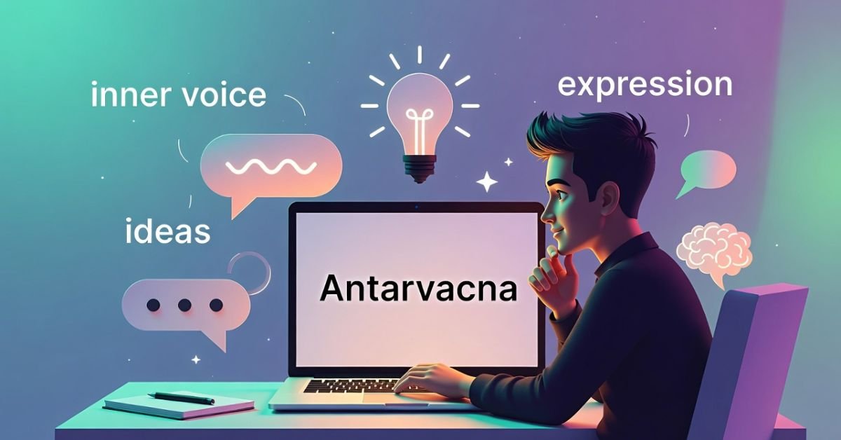
Antarvacna is composed of two Sanskrit roots: antar (अन्तर), meaning “inner” or “within,” and vacna (वाचन), meaning “speech,” “voice,” or “utterance.” Together, they describe something beautifully precise: inner speech the ongoing conversation you have with yourself at the deepest levels of awareness.
But Antarvacna is more than the idle chatter of an anxious mind. As a structured practice, it is the art of deliberately entering into dialogue with your own consciousness observing thoughts and emotions without identification, asking penetrating questions, and receiving the kind of clarity that only genuine self-inquiry can provide.
Think of it not as passive daydreaming, but as an active relationship with your inner world. Just as a skilled therapist asks questions that illuminate what a client could not see on their own, Antarvacna trains you to become that wise, compassionate witness for yourself.
Sanskrit Etymology
Antar (अन्तर) inner, within, between · Vacna (वाचन) speech, voice, expression · Together: the voice that speaks from within; the practice of conscious inner dialogue.
Antarvacna vs. Meditation vs. Mindfulness: What Is the Difference?
A common source of confusion is how Antarvacna relates to meditation and mindfulness. These concepts are deeply connected, yet each plays a distinct role in the journey of self-awareness. Understanding the difference is the first step to practising Antarvacna effectively.
| Practice | What it is | Primary focus | Relationship to Antarvacna |
|---|---|---|---|
| Mindfulness | Present-moment, non-judgmental awareness | Noticing what is sensations, thoughts, surroundings | Creates the calm clarity that makes Antarvacna possible |
| Meditation | A training tool for the mind | Focusing, relaxing, or observing the mind itself | Builds the mental stillness in which inner voice emerges |
| Antarvacna | Active inner dialogue and self-inquiry | Questioning, understanding, and transforming through inner conversation | Uses the stillness created by both to go deeper |
In short: mindfulness notices, meditation trains, and Antarvacna asks. It is the practice that takes the clarity cultivated in quiet sitting and transforms it into genuine self-knowledge and purposeful action.
OriginsThe Ancient Roots: History and Origins of Antarvacna
A Journey Through Ancient Indian Philosophy
The concept of Antarvacna does not exist in isolation. It emerges from one of the richest philosophical traditions on earth the ancient Indian pursuit of Atma-Vichara, or self-inquiry. The sages of the Upanishads, the foundational texts of Vedantic philosophy composed roughly between 800 BCE and 200 BCE, were fundamentally concerned with a single, burning question: Who am I, truly?
This was not abstract philosophy for its own sake. The sages understood, millennia before modern psychology confirmed it, that the unexamined mind is a source of suffering. Habitual thought patterns, unquestioned beliefs inherited from society and family, and the relentless noise of the ego all obscure what they called the Atman the true, unchanging self beneath the surface.
Antarvacna was the practical vehicle for this inquiry. Where formal meditation techniques aimed to quiet the mind entirely, the inner voice practice engaged that quiet mind in purposeful dialogue asking it to reveal its own depths, to surface what was hidden, and to guide the practitioner toward authentic living.
The Evolution of a Timeless Practice
What is remarkable about Antarvacna is how its core wisdom has endured. The specific techniques have evolved, been adapted by different schools of thought, and absorbed influences from Buddhist philosophy, Sufi contemplative traditions, and even contemporary psychology but the essential insight remains unchanged: you carry, within yourself, the answers to your most important questions.
In our hyper-connected age, where external opinions and digital voices compete loudly for our attention, the practice of going inward feels almost countercultural. And yet its resurgence in interest suggests that something in us recognises what it has lost and is seeking to recover it.
“The quieter you become, the more you are able to hear what has always been speaking.”

PrinciplesThe Core Principles: How Antarvacna Works
Antarvacna rests on three interlocking principles. Together, they form a complete framework for genuine inner dialogue one that goes far beyond simply “thinking about your feelings.”
Principle One Cultivating the Observer: Witnessing Your Thoughts
The first and most foundational principle is the development of what contemplative traditions call witness consciousness the capacity to observe your own thoughts and emotions without being swept away by them.
Most of us live in almost complete identification with our thoughts. When a worried thought arises “I’m going to fail this” we do not experience it as a thought passing through awareness. We experience it as truth, as who we are. Antarvacna begins the moment you create even a hair’s breadth of distance between the observer (you) and the observed (the thought).
This is not detachment. It is the opposite of numbness. By stepping back enough to watch your thoughts, you actually feel them more clearly, understand them more fully, and are no longer enslaved by them. You become the sky rather than the weather.
Principle Two Decoding Your Inner Voice: Intuition vs. Fear
Once you can observe your inner world with some degree of stillness, you encounter a challenge that every practitioner faces: the inner voice is not a single, unified signal. It is a chorus and not all voices in that chorus deserve equal attention.
Learning to distinguish genuine intuitive guidance from the noise of fear, anxiety, ego, and social conditioning is perhaps the most valuable skill Antarvacna develops. Here is a working framework:
| Quality | Intuitive Inner Voice | Fear-Based Inner Voice |
|---|---|---|
| Tone | Calm, clear, often quiet | Urgent, loud, insistent |
| Content | Pointing toward; expansive | Warning away from; contracting |
| Timing | Often arrives unbidden, in stillness | Tends to spiral and repeat |
| Feeling | Neutral clarity, even about difficult truths | Anxious, tight, catastrophising |
| Persistence | Returns quietly, consistently | Changes with mood and circumstance |
Antarvacna does not suppress the fear-based voice it listens to it with compassion, understands its protective intent, and gently inquires: Is this true? Is this serving me? What does this fear actually need?
Principle Three Dialogue and Inquiry: Asking the Right Questions
The third principle transforms passive observation into active self-inquiry. Antarvacna is a practice of asking and the quality of your questions determines the quality of the insights you receive.
Some of the most powerful self-inquiry questions used in the practice include: What is this thought actually trying to protect me from? What would I do if I were not afraid? What truly matters to me beyond what I have been taught to want? What am I avoiding looking at?
These are not rhetorical questions to be answered quickly. They are invitations for the deeper intelligence within you to respond often through a felt sense of recognition, a shift in perspective, or a quiet knowing that gradually grows clearer over days of practice.
BenefitsThe Transformative Benefits of Practising Antarvacna
The benefits of a consistent Antarvacna practice are not abstract promises they are the natural consequences of genuine self-knowledge. Here are the four most significant transformations practitioners report.
Enhanced Self-Awareness & Emotional Intelligence
As you learn to witness your inner world, you develop a nuanced understanding of your emotional patterns, triggers, and motivations. This is the foundation of emotional intelligence not managing emotions, but understanding them.
Sharper Decision-Making & Clarity
When you know your authentic values from the inside out rather than the outside in, decision-making becomes less agonising. You have a reliable inner compass to consult when external opinions conflict.
Improved Mental Health & Stress Reduction
Research on journaling and introspective practices consistently shows reductions in stress and anxiety. Processing emotions through Antarvacna prevents them from accumulating as unexamined inner pressure.
Accelerated Personal Growth & Fulfilment
Living in alignment with your authentic self rather than the self shaped entirely by external expectations produces a deep sense of purpose and meaning that ambition alone cannot provide.
PracticeA Step-by-Step Guide: How to Practise Antarvacna Daily
This is the most important section of this guide. Antarvacna is not an idea to admire from a distance it is a practice to do. Here is a complete framework for beginning and sustaining your practice, regardless of your schedule or experience.
Preparing Your Space and Mindset
You do not need elaborate rituals or special equipment. What you need is five to twenty minutes of genuinely uninterrupted time and a space where you feel safe. A consistent, dedicated space even a particular chair signals to your nervous system that this is time for inward attention.
Before beginning, set a simple intention. Not a goal to achieve, but a direction to face: I am here to listen honestly to myself. This small act of deliberate orientation makes a meaningful difference.
The 10-Minute Foundational Practice
Settle and Breathe 2 minutes
Sit comfortably, close your eyes, and take three slow, deliberate breaths. Allow your exhales to be noticeably longer than your inhales this activates the parasympathetic nervous system and signals safety to your body. Let the first two minutes simply be about arriving.
Observe Without Judgement 3 minutes
With eyes still closed, simply notice what is present in your inner world. Thoughts, emotions, physical sensations, background feelings. Do not try to change anything. Practise being the witness: There is worry. There is tiredness. There is a background tension in my shoulders. Name what you find, then watch it.
Inquire Gently 3 minutes
Choose one thing that surfaced in the previous step perhaps an emotion, a recurring thought, or a tension and ask it a single, open question. What are you trying to tell me? What do you need? Where did you come from? Do not force an answer. Sit with the question and notice what arises images, feelings, words, or simply a shift in your inner atmosphere.
Journal Your Insights 2 minutes
Before leaving your practice, write two or three sentences capturing what emerged. This step is more important than it seems it externalises the insight, making it more real and easier to return to. Over weeks and months, your journal becomes an extraordinary map of your inner landscape.
Advanced Techniques for Deeper Exploration
Once the foundational practice feels natural, these techniques open new dimensions of self-inquiry:
Guided Visualisation
Imagine a wise, compassionate version of yourself future or symbolic and hold a conversation with them about your current situation. Ask what they see that you cannot.
Walking Contemplation
Take a 20-minute walk in nature with a single question held lightly in mind. Do not seek an answer actively allow the rhythm of movement to loosen insights that stillness could not.
Expressive Writing
Write continuously for 10 minutes from the perspective of an emotion or situation: “I am your fear of failure, and here is what I need you to know…” The perspective shift unlocks remarkable honesty.
Integrating Antarvacna into a Busy Schedule
The full practice described above requires dedicated time but the spirit of Antarvacna can be woven throughout an ordinary day through what practitioners call micro-practices:
- Mindful commute:Instead of reaching for your phone, sit with a single open question for the first five minutes of your journey.
- Pre-meeting pause:Before entering an important conversation or decision, take three breaths and ask: “What do I actually feel about this situation?”
- Evening review:Spend three minutes before sleep asking: “What surprised me about myself today? What did I avoid?”
- Transition moments:The spaces between activities standing in a queue, waiting for a file to load are micro-opportunities for brief inner listening.
- The one-question practice:Choose a single self-inquiry question each morning and carry it with you, gently returning to it throughout the day.
ChallengesOvercoming Common Challenges on the Path of Antarvacna
Every sincere practitioner encounters obstacles. Here are the most common, and how to work with them rather than against them.
“I can’t hear my inner voice there’s just noise.”
This is almost universal in the beginning. The inner voice has not disappeared it has simply been crowded out by the relentless stimulus of modern life. Before attempting inquiry, spend more time in the observation phase. Do a brief body scan: notice sensations from head to feet. This anchors awareness in the present and gradually creates the space in which subtler signals can surface. Start with five minutes. Consistency matters more than duration.
“What I find inside is uncomfortable even frightening.”
This is a sign the practice is working. Antarvacna does not promise only pleasant discoveries. Unacknowledged grief, buried anger, long-suppressed desires these often surface when we finally create the conditions for honest self-encounter. Approach difficult material with the same compassionate curiosity you would offer a friend in pain. If the material feels overwhelming, it is wise to work with a therapist alongside your practice there is no contradiction between ancient wisdom and modern professional support.
“I practise for a few days and then forget entirely.”
The enemy of any contemplative practice is not resistance it is abstraction. Tie your Antarvacna practice to an existing daily anchor: immediately after morning coffee, before brushing teeth at night, or at the very start of your lunch break. The habit architecture of “after X, I do Y” is far more reliable than willpower alone. And when you miss days which you will begin again without judgment. The capacity to return is itself part of the practice.
॰ ✦ ॰
Frequently Asked Questions About Antarvacna
Is Antarvacna a religious practice?
Antarvacna has roots in ancient Indian philosophical traditions, but it is not a religious practice in the devotional sense. It requires no particular beliefs, rituals, or affiliation. People of all faiths and none practise variations of inner dialogue and self-inquiry. What it does require is sincerity and a genuine willingness to look honestly at oneself.
How is Antarvacna different from psychotherapy?
Both pursue self-understanding, but through different means. Psychotherapy is a clinical relationship with a trained professional who brings external expertise, diagnostic frameworks, and structured interventions. Antarvacna is a self-directed practice of inner inquiry. The two are deeply complementary many therapists incorporate self-inquiry techniques, and many Antarvacna practitioners find that therapy accelerates what self-practice begins.
How long does it take to see results?
Many practitioners notice shifts in clarity and emotional awareness within two to three weeks of consistent daily practice even ten minutes a day. Deeper transformations in values-alignment, habitual patterns, and life direction emerge over months. The practice has a compounding quality: each session builds on the self-knowledge accumulated in previous ones.
What is the best time of day to practise?
Early morning is traditionally preferred the mind is quieter before the day’s demands have accumulated, and insights gained in the morning can be carried consciously through the day. Evening practice offers the advantage of processing the day’s experiences while they are still fresh. The honest answer is: the best time is the time you will actually do it consistently.
How do I know if I’m hearing intuition or just my ego?
This is one of the most valuable distinctions Antarvacna develops over time. As a general guide: intuition tends to be quiet, consistent, and neutral in tone it returns reliably, even when you try to dismiss it. The ego tends to be loud, reactive, and emotionally charged it argues, justifies, and changes its position based on fear and desire. The witness practice of Antarvacna gradually trains this discernment through direct experience rather than theory.
Can Antarvacna help with anxiety and depression?
Self-inquiry and introspective journaling practices have well-documented benefits for psychological wellbeing. Processing emotions through structured inner dialogue can significantly reduce the burden of unexamined rumination. However, Antarvacna is a self-development practice, not a clinical intervention. If you are experiencing clinical anxiety or depression, please seek support from a qualified mental health professional. The two approaches can work in powerful conjunction.
-

 TECH9 months ago
Apple iPhone 17: Official 2025 Release Date Revealed
-

 BLOG9 months ago
Understanding the ∴ Symbol in Math
-

 ENTERTAINMENT7 months ago
What Is SUV? A Family-Friendly Vehicle Explained
-

 EDUCATION4 weeks ago
Horizontal Translation: How to Shift Graphs
-

 EDUCATION9 months ago
Using the Quadratic Formula
-

 ENTERTAINMENT9 months ago
Going Live: How to Stream on TikTok from Your PC
-

 TECH9 months ago
Prodigy: Early Internet Pioneer
-

 EDUCATION9 months ago
The Meaning of an Open Circle in Math Explained